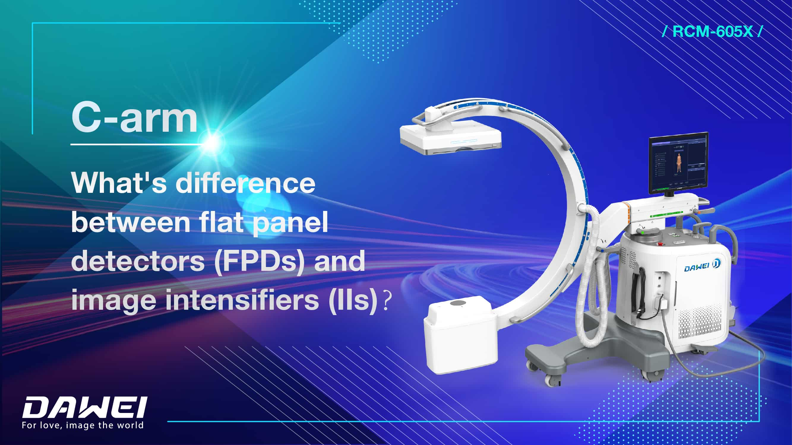
The Great C-Arm Dilemma: Image Intensifier vs. Flat Panel Detector – A Definitive Guide for Medical Practitioners
Introduction: The Evolution of Real-Time Imaging
C-arm systems revolutionized medical imaging by providing real-time X-ray visualization during surgeries, pain management procedures, and interventional diagnostics. At the heart of these systems lies a critical technological choice: traditional image intensifiers (II) or modern flat panel detectors (FPD).

1. Core Technology: How They Transform X-Rays into Images
Image Intensifiers (1950s Technology):
X-rays strike an input phosphor, converting them to electrons. These electrons are accelerated through a vacuum tube, strike an output phosphor, and create a visible light image captured by a camera. This multi-step analog process introduces geometric distortion (especially at image edges) and progressive gain degradation due to phosphor wear.
Flat Panel Detectors (1990s Technology):
X-rays strike a scintillator layer (cesium iodide or gadolinium oxysulfide), converting them to visible light. This light is immediately captured by an amorphous silicon photodetector array and converted directly into a digital signal. This streamlined process eliminates distortion and maintains consistent image quality over time.
2. Critical Comparison: Performance and Operational Impact
Feature | Image Intensifier (II) | Flat Panel Detector (FPD) |
Image Resolution | Moderate (degrades over time) | High (consistent, up to 2k X 1) |
Geometric Distortion | Significant ( up to 10-15% edge errors) | Minimal (≈1%) |
Field of View | Limited | Larger |
Radiation Dose | Higher (5x increase in Zoom 3 mode) | Lower (more efficient detection) |
Lifespan Consistency | Degrades after 5-7 years | Stable>10 years |
Physical Size | Bulky tube design | Slim profile |
Heat Generation | Significant | Minimal |
3. Clinical Applications: Matching Technology to Specialties
Clinical Applications | Image Intensifier (II) | Flat Panel Detector (FPD) |
Orthopedics & Vascular Surgery |
| FPDs dominate due to higher resolution |
Pain Management & Basic Fluoroscopy | viable for lower-resolution needs |
|
Pediatrics & High-Dose Specialties |
| FPDs’ dose efficiency of less than 30-50% lower exposure is critical for patients and long procedures. |
FPDs are phasing out IIs as manufacturing costs drop and software capabilities expand. Emerging trends favor FPDs:
AI-driven image enhancement and dose management
Miniaturization for point-of-care imaging
Integration with robotic surgical platforms
Refurbished IIs will persist in price-sensitive markets, but FPDs are becoming the clinical standard.
English
العربية
Français
Русский
Español
Português
Deutsch
italiano
日本語
한국어
Nederlands
Tiếng Việt
ไทย
Polski
Türkçe
ພາສາລາວ
ភាសាខ្មែរ
Bahasa Melayu
ဗမာစာ
Filipino
Bahasa Indonesia
magyar
Română
Čeština
Монгол
қазақ
Српски
हिन्दी
Slovenčina
Slovenščina
Norsk
Svenska
українська
Ελληνικά
Suomi
Հայերեն
עברית
Latine
Dansk
Shqip
বাংলা
Hrvatski
Afrikaans
Gaeilge
Eesti keel
Māori
नेपाली
Oʻzbekcha
Български
ქართული
Кыргызча


